
This sample Forensic Anthropology Research Paper is published for educational and informational purposes only. If you need help writing your assignment, please use our research paper writing service and buy a paper on any topic at affordable price. Also check our tips on how to write a research paper, see the lists of criminal justice research paper topics, and browse research paper examples.
Overview
Forensic anthropology, a subdiscipline of physical anthropology, is dedicated to the biological identification of human remains discovered within a legal context in advanced stages of decomposition, or in situations where the integrity of the body has been compromised, thereby thwarting a positive identification through conventional means. Since its inception during the late nineteenth century (Stewart 1979), forensic anthropology has matured as a science, and professionals in this field have become actively involved in domestic or international police investigations, human rights investigations, and disaster victim identification (DVI), forming an integral element of body handling within the mortuary and in some instances in the field during the search and location of victims.
The advent of global participation and communication among forensic anthropologists has led to the development of new methodologies, such as state-of-the-art 3D imaging technology, which assist with the biological identification of human remains within a variety of contexts and circumstances.
This research paper aims to present the historical process from which the discipline has evolved, and sheds light upon the application and methods utilized when a biological identification is required during a legal investigation. Current challenges are expanded upon; the future of the discipline is discussed and also potential avenues for future research.
Fundamentals Of Forensic Anthropology
Developmental Background
The origins of forensic anthropology commenced within the United States during the late nineteenth century, in connection with studies conducted by the anatomist T. Dwight (1843–1911), concerning among many elements, the determination of sex, age, stature, and variability in relation to the human skeleton; his contribution led to the title of Father of American Forensic Anthropology to be conferred upon him (Stewart 1979). Dwight’s research constituted a catalyst for this discipline, and also served to inspire those who followed. When viewed in relation to modern standards, his studies may appear rather basic, but they provided the initial impetus for the promotion of a discipline that has aided law enforcement organizations with the identification of human remains through many decades and, within a more recent context, involvement in trauma identification and description related to cause and manner of death. Dwight’s studies were continued by the American anthropologist G. Dorsey (1868–1931) into the early part of the twentieth century, at which time the latter offered a significant contribution to the development and refinement of techniques applied for human identification based on bone analysis; in addition, his intervention as an expert witness gave the discipline an initial profile within the crimninal justice system (Klepinger 2006; Stewart 1979).
The twentieth century can be seen as the era in which engagements with law enforcement agencies and the necessity to identify war casualties constituted decisive junctures which led to forensic anthropology becoming a valid discipline in its own right. During the last century, a particularly important figure which stands out is A. Hrdlicˇka (1869–1943). Hrdlicˇka, who was formally trained in medicine, was director of the Anthropology Department at the Smithsonian Institution, where he amassed an impressive collection of human remains, and conducted active research in the field of physical anthropology. Additionally, Hrdlicˇka was the founder of the American Journal of Physical Anthropology (1918) and also the American Association of Physical Anthropologists (1930), of which both elements aided in providing a formal grounding to this discipline; within the area of forensic intervention, Hrdlicˇka received skeletal cases from the Federal Bureau of Investigation (FBI) (Ubelaker 1999). Parallel to the work of Hrdlicˇka was that of W. Krogman (1903–1987), who in 1939 published “Guide to the Identification of Human Skeletal Material” in the FBI Law Enforcement Bulletin, considered by some as the publication that formally recognized the specific importance of forensic anthropology (Tersigni-Tarrant and Shirley 2013; Stewart 1979; Ubelaker 1999). With the advent of Krogman’s publication, a guide was available that provided a point of reference for further research, in addition to constituting a valuable reference guide for use in the field.
Upon Hrdlicˇka’s death, T. Dale Stewart (1901–1997) took over the post at the Smithsonian Institution; not only were investigative cases emanating from the FBI continued, but the valuable contributions that Stewart made to academic teaching and research improved and promoted new developments in bone analysis within forensic cases (Kennedy 2000).
In 1972 the physical anthropologist E. Kerley (1924–1998) founded the Physical Anthropology Section in the American Academy of Forensic Sciences (AAFS), and it is here where those involved in the analysis of human remains within the legal context were recognized formally within a prestigious association. This advance was followed by the possibility of members being formally certified through the American Board of Forensic Anthropology (ABFA), an organization created to foster and maintain standards within the profession and to confer professional weight and credentials to those practicing members (Tersigni-Tarrant and Shirley 2013).
These important events led to more in-depth research and initiatives, which were further strengthened when the US armed forces recognized the requirement for the identification of human remains of those who perished in combat. Such interventions occurred in World War II (1939–1945) and the Korean War (1950–1953) and during the Vietnam War (1965–1975) (Stewart 1979; Klepinger 2006), a challenge that has increased exponentially in relation to advancements in weapons technology. As a result of such interventions, the US Army Central Identification Laboratory, Hawaii (CILHI), was established in 1976; today it is called Joint POWMIA Accounting Command (Joint Prisoners of War Missing in Action Accounting Command) (JPAC 2013), with the mission of locating all of the Americans who are still missing from past wars. This is achieved by locating, recovering, and identifying the remains, a process whereby forensic anthropology is a key component for a successful outcome (JPAC 2013; Tersigni-Tarrant and Shirley 2013).
As a result of its evolution with respect to these major events, forensic anthropology became established as a specialized forensic field in its own right.
Definition And Functions
Forensic anthropology can be defined as a subdiscipline of physical anthropology (also referred to as biological anthropology), which conducts biological identifications on human remains within a legal context and in circumstances when they are in a poor state of preservation including advanced stages of decomposition, dismemberment, fragmented or possibly incomplete, burnt, skeletonized, saponified, or a combination of such factors (Nafte 2000; Klepinger 2006) (see Figs. 1 and 2). Within such scenarios, saponification occurs when the body, whole or in part, is covered with adipocere, a grayish waxlike substance formed by the hydrolysis of body fats when exposed to an environment which is moist and low in oxygen, such as the case with bodies held underwater; however, saponification may also occur in desertlike environments when the water content in the fat cells compensates for the lack of moisture within the environment (Gill-King 1997).
The process of identification is accomplished via biological profiling of the body in order to determine sex, age, stature, biological affinity (ancestry), and individual characteristics through bone analysis. In some individual cases, the forensic anthropologist will also be involved with trauma identification on bone in instances such as sharp (see Fig. 3) and blunt force trauma, gunshot wounds (see Fig. 4), strangulation and in circumstances in which a possible interpretation of events is required, such as cases where there is a requirement to determine if the event was peri-mortem (around the time of death) or postmortem (Byers 2008; Klepinger 2006; White and Folkens 2005). If the remains in question retain soft tissue of any kind, the forensic anthropologist will, as a matter of course, macerate the remains in order to access the bone tissue. However, in relation to the latter scenario, new and less invasive approaches through 3D imaging techniques are currently being developed (see Current Challenges and Future Directions section).
The profiling process is based on applying metric and nonmetric (morphological characteristics) assessment when appropriate, as both approaches are normally utilized in a complementary fashion. Ideally, all of the potential methodologies that can be applied should be used, as this will increase the possibility of a positive outcome.
- Sex determination is normally the first step taken when producing a biological profile of human remains, as this element is also linked to the estimation of ancestry and the determination of stature (refer to appropriate sections below).
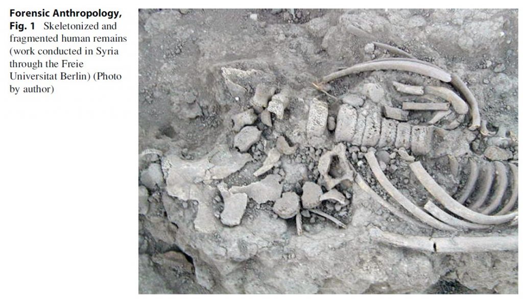 Forensic Anthropology, Fig. 1 Skeletonized and fragmented human remains (work conducted in Syria through the Freie Universitat Berlin) (Photo by author)
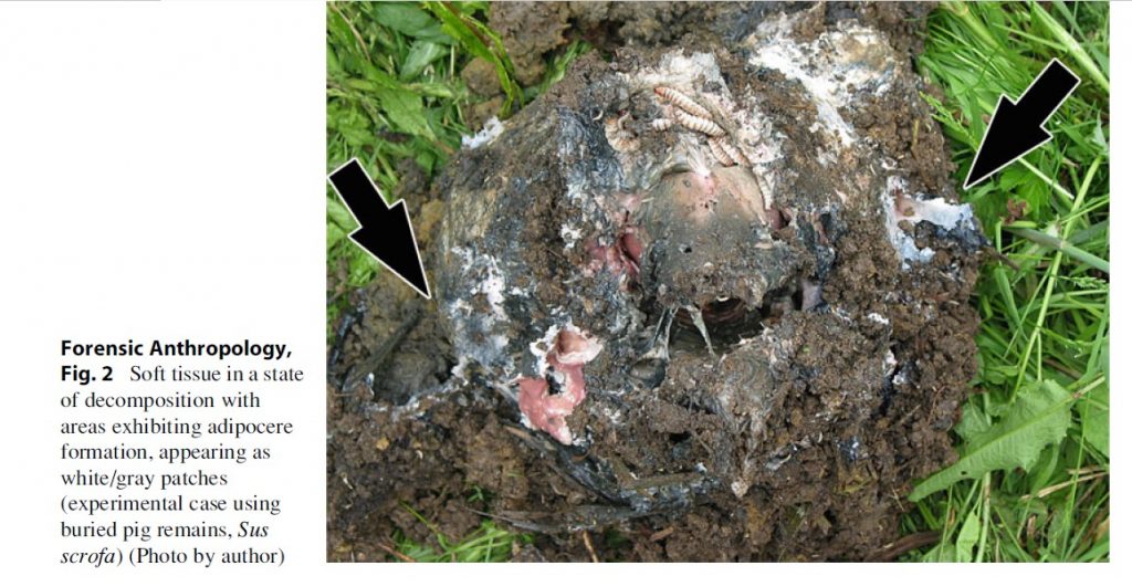 Forensic Anthropology, Fig. 2 Soft tissue in a state of decomposition with areas exhibiting adipocere formation, appearing as white/gray patches (experimental case using buried pig remains, Sus scrofa) (Photo by author)
Determination of sex may be arrived at by morphological and metric analysis of adult skeletal remains (defined as those with finalized bone and dental development and which exhibit secondary sexual changes). Remains belonging to juveniles, also referred to as adolescents (characterized when secondary sexual changes are taking place) or younger individuals, are not normally sexed; in such cases, the skeleton is still going through a process of development, and sexually dimorphic characteristics (sexual dimorphism being the difference in size and shape between males and females) have not fully developed (Scheuer and Black 2000; Nafte 2000); when attempts at the determination of sex in adolescents are made, the results generally tend to be weak. Another consideration within the determination process is that traits may vary somewhat from one population to another, with some being more dimorphic than others; it is through accumulated experience that a forensic anthropologist gains insight into the specifics evident within any given group.
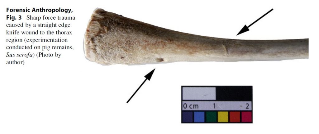 Forensic Anthropology, Fig. 3 Sharp force trauma caused by a straight edge knife wound to the thorax region (experimentation conducted on pig remains, Sus scrofa) (Photo by author)
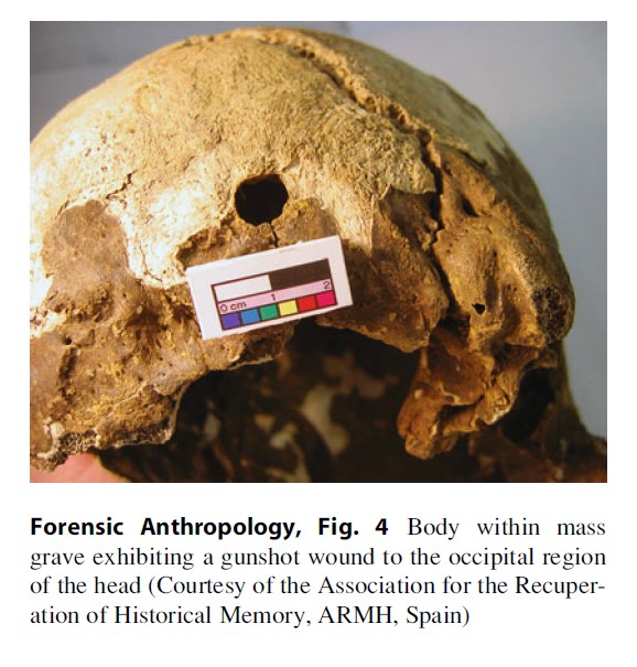 Forensic Anthropology, Fig. 4 Body within mass grave exhibiting a gunshot wound to the occipital region of the head (Courtesy of the Association for the Recuperation of Historical Memory, ARMH, Spain)
Various individual bones have been recently studied with the aim of developing new techniques pertaining to sex determination, with some approaches producing better results than others; however, the pelvis and the skull are still considered as the two areas of the body which are most reliable in this respect. In the case of the pelvis, composed of a sacrum and a right and left os coxa (hip bone), analysis through the Phenice’s method, a morphological assessment of the pelvis, is reported to be as accurate as 96–100 % (Silva Braz 2009; Mays 2010), as this structure possesses well-defined sexually dimorphic traits associated with the ability of childbearing in females.
With reference to the skull, the major attributable differences are due to males being larger and with more robusticity, translating into noticeable muscle insertions upon bone, with competent analysis producing a rate of 92 % accuracy (Mays 2010).
Conversely, the metric approach uses a series of measurements on specified landmarks on bone. When using discriminant functions, the results are fed into a set formula that provides an ending score, which is subsequently compared against the sectioning point (determining the sex based upon the score obtained). A problem that is frequently encountered with available formulas is that they are population based, and as such, the results can be skewed if the remains do not belong to the relevant database. Moreover, the metric analysis can also be inputted into the computerized program Fordisc 3 (FD3), based on contemporary data gathered from the Forensic Data Bank at the University of Tennessee in Knoxville, USA (Sauer and Wankmiller 2009; TersigniTarrant and Shirley 2013). The negative side of using FD3 is that the said database is also population specific, with the possibility of the same problems arising as mentioned above.
- Age determination is a step that is applied to subadult and adult remains alike, in order to arrive at the age of death through morphological observations and metric analysis applied to dental and bone remains.
– Subadults – subadult remains offer a wealth of information as the growth development follows a set sequence of events at the bone and dental level. These parameters have been cross-referenced with age and have been converted into what are referred to as growth standards, which are used by the forensic anthropologist in comparison with observations made on the final growth stage evident upon the remains being examined (Nafte 2000; Klepinger 2006). Because these processes of development tend to be standard in nature across populations, the overall results are highly accurate.
Dental development may be appreciated through tooth mineralization and eruption and constitutes the most accurate method for aging as it is a constant and continuous process which has been well documented. Dental remains may be examined individually if found dislodged from the alveolar process (the socket which holds each dental piece in the upper and lower mandibles); furthermore, in cases when teeth have not erupted, they may be analyzed through x-rays or extracted and examined microscopically (Klepinger 2006). In the latter case, it is possible to determine if an infant was stillborn or a live birth as evidenced by the neonatal line, located upon the enamel surface on all deciduous dentition and first permanent molars, formed at birth or soon afterwards (Scheuer and Black 2000).
Bone development can be assessed in subadults, and in the case of fetal remains, the bones can be measured from prescribed landmarks and compared with established growth standards that indicate the gestational age with relatively good confidence levels (Scheuer and Black 2000; Fazekas and Ko´ sa 1978). It is after birth, however, that intrinsic and extrinsic factors play a strong role in skeletal development, creating variations in developmental rates at individual and population levels. As indicated by Byers (2008), age assessment in relation to the length of long bones may be applied up until the age of 10, at which time the correlation between age and length starts to widen, producing inaccurate age estimations.
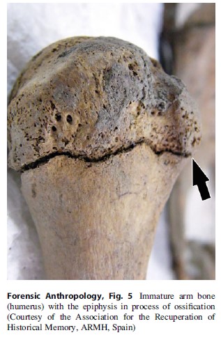 Forensic Anthropology, Fig. 5 Immature arm bone (humerus) with the epiphysis in process of ossification (Courtesy of the Association for the Recuperation of Historical Memory, ARMH, Spain)
Another method utilized to establish age at death is through the identification of primary ossification centers (areas where bone is deposited), as well as their union with other primary centers or with secondary ossification centers (see Fig. 5). Because of their small size, the identification of primary centers may prove difficult to recognize and recover at a scene when dealing with skeletonized remains or bodies in which integrity has been compromised; this method is best employed in connection with examination of the skull, mandible, upper cervical vertebrae, and ribs. On the other hand, the union between the primary and secondary centers is frequently more productive within forensic anthropology, as they unite at a scheduled rate (as indicated earlier with individual variations and population trends) extending into early adulthood; such is the case of the clavicle, in which final fusion ends at approximately 29 years of age (Scheuer and Black 2000; White and Folkens 2005).
– Adults – in the case of adult remains, age is determined by morphological changes visible through the naked eye, which in some instances may be degenerative in nature, and by remodeling processes at the microscopic level. The main areas within the skeleton that aid in age determination through morphological changes are the pubic symphysis, the auricular surface of the ilium upon the os coxa, the sternal end of the fourth right rib, and the cranial sutures (White and Folkens 2005; Byers 2008; Klepinger 2006). Additionally, it is also possible to obtain an estimate of chronological age through dental analysis of the incisors, canines, and premolars as specified by the Lamendin’s technique.
In the case of the first three skeletal areas, these are widely used, as a process of “metamorphosis” or changes in the bone morphology occurs progressively as a person ages, associated with sex, and also to a degree with respect to population affiliation; these changes possess set characteristics which are compared against a set of representative age stages. In the case of the pubic symphysis, various methods are available including the Suchey-Brooks model (White and Folkens 2005), which is widely utilized as it constitutes a multiracial database, but is, however, not wide enough to encompass all populations; as such, problems have been reported in the past in connection with skewed results in relation to populations that are not represented within its database.
With regard to cranial sutures, the skull is examined at specific points in order to assess the degree of closure, whereby a prescribed value is scored for each area examined; these values are then compared against an existing table to obtain an age range. Because closure is not a linear occurrence associated with age, the results may indicate wide age ranges making this method somewhat unreliable, such as within cases in which only the skull is recovered without dental remains, or present in poor condition and not suitable for ageing.
Dental remains are normally utilized with relation to subadults in order to obtain a chronological age at death in association with the developmental stage in evidence; however, the Lamendin’s dental aging technique was designed to more accurately determine the age of adult dental remains. The latter technique consists of examining single rooted teeth based upon their degree of root translucency and the degree of periodontosis (recession of the gum line). Research indicates that results are linked to sex and, to a lesser degree, to ancestry; however, this method may not be desirable if applied to young adults. Results may also be influenced by additional variables such as dental hygiene and dental treatment intervention (Prince and Ubelaker 2002).
Finally, the histological method consists of examining changes in bones and the teeth at a microscopic level; however, this method is time consuming and requires the use of specialized equipment and specific training (Byers 2008), and as such it is not always a viable option in some cases.
- Biological affinity or ancestry is the most difficult task that a forensic anthropologist attempts to evaluate when biologically profiling human remains, producing results which may be somewhat tenuous; however, the estimation of ancestry within an investigation is a necessary element, as it provides an avenue of inquiry for the authorities. Additionally, this information is also required when calculating stature (see section on Stature).
More specifically, the challenge arises from the element of phenotypic traits (expression of the genetic makeup) reflected at the skeletal level in one given population being small in number, but which may also be shared in part by other populations with added variation, thereby creating the possibility of a continuum wherein boundaries are blurred, not to mention the inherent variability that exists at the interpopulation level. Furthermore, sex-determining traits can influence the traits associated with estimating ancestry (Klepinger 2006). Additionally, individuals within any given population may describe or view themselves not in biological terms but by device of ethnic/social classification. For example, many Latin Americans would describe themselves as white or Hispanic; however, their morphological traits would not necessarily be categorized as white, and Hispanic is a term stemming from an ethnic category, something that cannot be verified biologically. To further confound this issue, the high level of migrant influx and intermarriages within any given geographical zone will intensify the challenges with respect to identifying any given question of biological affinity.
At present, global populations are divided into different groups, deriving from genetic or skeletal research, whether referring to the global population as a whole or to a specific population within a particular country. At the skeletal level, four main ancestry categories have been created: Caucasoid (white American/European) (white American/ European), Mongoloid (East Asian), Negroid (African black), and Native American (Byers 2008; Klepinger 2006; Tersigni-Tarrant and Shirley 2013). As it may be appreciated, this categorization does not embrace all of the global geographical regions.
Attempts to assess ancestry through traits present on bone are accomplished primarily through morphological or metric analysis of the craniofacial region, as this is where traits are most effectively expressed, mainly with reference to the shape of the head and nasal region and additionally the upper mandible. Within a given morphological analysis, a sum of characteristics is sought and subsequently compared to known samples to arrive at a determination (Byers 2008; Klepinger 2006), a task best accomplished by an experienced anthropologist. Because the estimation is based upon morphological observations, and not all traits may be strongly reflected within one particular individual, the results obtained may be argued over due to the element of subjectivity. Conversely, a metric analysis compiled between specific landmarks can be perceived as a more accurate, objective approach.

Metric analysis can also be fed into the FD3 software, which contains data for this purpose drawn from contemporary remains from the USA, in addition to world samples, and representative of various archaeological populations (Tersigni-Tarrant and Shirley 2013); as well, another computer programme available to forensic anthropologists is CRANID, with a data base comprised of 74 samples (3.163 skulls) from various regions of the world (Sauer and Wankmiller 2009; Wright 2012). However, the application may not provide adequate results when the affinity of the specimen in question has not been represented within the available databases.
Postcranial bones may be used, with the femur (leg bone) being considered as being the best choice as it possesses various traits and has been comprehensively studied (Byers 2008; Sauer and Wankmiller 2009). However, in general terms, the postcranial bones retain fewer traits which are not as clearly expressed as in the craniofacial region, thereby reducing confidence levels with respect to accuracy.
Research has been attempted upon dental material, but this particular avenue of inquiry has not been as comprehensive as that completed with respect to bone; additionally, dental traits are fluid, varying in degree, and not strictly present within any given population.
For the reasons stated herein, it is always recommended to collate results from all available methods and to be actively aware that a definite determination of ancestry from the remains is not always possible, but predominantly an educated estimation.
- Stature – calculating the stature at the time of death is accomplished by measuring a selected long bone from the upper or lower extremities, with the result computed into a regression formula that has been designed by bone type, sex, and biological affinity; a proportional correlation may then be compared between the bone used and the height of the unknown individual. The result obtained will constitute the mean height with a standard deviation. The femur and tibia are the most widely utilized reference points, as the results obtained from these bones provide more accurate outcomes (Byers 2008).
In cases in which the body is highly incomplete or fragmented, such as when dealing with dismemberment, air crashes, or the victims of terrorist acts, there are a series of methods available that allow estimation of the stature of any given individual through the use of small segments from the long bones (Byers 2008). Because of the nature of these measurements, the results are not as accurate, but are still helpful in cases where an approximation of height is required. When skeletal remains are dry, due to time lapse, shrinkage can occur, and adjustments can be necessary in order to compensate for such factors through pre-established data; the same approach may be taken with remains that are determined to be over 45 years of age, as stature starts to diminish beyond this period (Byers 2008). It should also be clearly stressed that all populations are not represented within all available formulas, and as such, adjustments must be made and considered when confronted with each particular case.
The results obtained from such calculations often prove difficult to verify, as accurate reference to stature is information that is not often available through family members and friends or via official records.
- Individual characteristics – individual traits can aid in an investigation for two reasons: when there are no indications of who it may be or when the authorities have a suspicion of who the person may be. Such traits may include evidence of a fracture or amputation that the person may have suffered in life, with time to heal through bone remodeling, including misalignment of segments which produce evidence of the event (Byers 2008).
Other traits include pathologies left on bone that can indicate past conditions, diseases, or habits, as in the case of osteoporosis upon the skull, associated with malnutrition and drug addiction (Klepinger 2006); an infected tooth may cause an abscess which produces a small opening on the jaw bone. Diseases may also affect bone, such as tuberculosis, a chronic infection caused by a bacteria which can leave marks in the vertebral column, and in other bones such the os coxae (hip bones). Furthermore, congenital anomalies due to improper formation can aid in the identification of a person and may be present in various parts of the body, such as the vertebral column, hands, and feet (Otner 2003; Klepinger 2006; White and Folkens 2005).
The human skeleton also possesses traits which can assist in the individualization process, as is the case of improper non-fusion of segments conforming a bone, missing bone sections, or characteristics within a particular bone which are not pathological in nature, but which can be identified and compared against an antemortem x-ray or computer tomography image (CT).
All of the above conditions may be used to guide investigations, and with cross-referencing the information gathered in tandem with medical records, which can aid in the identification process.
Facial reconstruction is a method which can aid in the identification process by applying a variety of methods from sculpting in clay to the use of sophisticated computer graphic programs (Wilkinson 2008); such avenues are attempted by individuals from a variety of disciplines, including those from purely artistic backgrounds to forensic anthropologists who specialize within this specific field.
The described steps that a forensic anthropologist takes in order to compile a biological identification can lead to a positive identification through DNA, odontological, and/or medical analysis, once the investigating personnel have a description with which to guide them.
Trauma Analysis
The determination of the cause and manner of death legally rests with the forensic pathologist; however, forensic anthropologists do participate within cases in which bone reconstruction is required due to fragmentation caused by high-velocity projectiles or in connection with blunt force trauma; in both instances, an interpretation and sequence of events can be indicated when possible, as in cases when more than one impact has been inflicted upon the skull. Interpretations concerning the circumstances in which the trauma was caused can be assessed; an example would be when, there is a need to determine whether an individual fell or jumped intentionally or to determine the angle of force in which a chest cavity was crushed, as would be evidenced in cases of severe stamping. Additionally, in the case of sharp force trauma, the type of weapon used, such as a knife (see Fig. 3), axe, hatchet, or machete, may be accurately identified, in addition to the angle with which the implement was used. In the case of dismemberment through sawing, the type of striations deposited upon the cut site can aid in a more specific identification of the tool which was utilized (Byers 2008; Galloway 1999; Nafte 2000).
In cases where strangulation was the cause of death, a close examination of the hyoid bone (located in the anterior side of the neck) may show signs of subtle breakage which can be interpreted by an experienced anthropologist.
With reference to children, this bone is very resilient as it has not ossified; hence breakage may not occur (Byers 2008; Klepinger 2006).
Depending upon the condition in which a body is discovered, including burnt remains, there may be a requirement for the anthropologist to determine if a given alteration on bone is peri-mortem or postmortem, a detail which may produce ramifications within a legal investigation; in some cases this element may be difficult to assess, necessitating specialized
Investigations Into Human Rights Violations
Investigations of human rights violations have given way to interventions in countries such as Guatemala, El Salvador, Colombia, Chile, Argentina, Democratic Republic of Congo, Rwanda, East Timor, Bosnia-Herzegovina, and Spain, where forensic anthropology has played an important part within multidisciplinary forensic teams. Among the many challenges encountered prior to deployment, one aspect that concerns work within forensic anthropology is to select the appropriate methods to be used as references for determining the biological profile of the victims, keeping in mind the genetic makeup of the population to be examined (as indicated above) (Ferllini 2007; EAAF 2013); as previously highlighted, inherent variations exist within populations and all references utilized should be directed at the required analysis in order to avoid the production of skewed results.
Within this arena, work is accomplished both within the field and at the mortuary. In the field, a comprehensive knowledge of bone anatomy is crucial during the search for human remains which may have been left on the surface and thereby exposed to environmental elements. It is also advantageous to be able to recognize small remains and distinguish them from animal bones within an outdoor setting. In situations when bodies are comingled and dispersed, an attempt is made to determine the number of individuals that are present while in situ, aiding in the implementation of proper lifting and bagging procedures prior to sending the remains to the assigned mortuary, which in some instances may be a temporary installation in close proximity to the area being surveyed.
In the case of buried remains, the forensic anthropologist is also directly involved with the exhumation process, whether dealing with the contexts of single or mass graves. With reference to the latter scenario, hundreds of bodies may be present, resulting from a single event, as was the case after the Rwandan genocide (1994), and where the remains may be comingled and skeletonized. In such cases the forensic anthropologist determines which bones pertain to any given individual by applying analysis of bone morphology and determining a general sex and age identity, elements which are verified at the mortuary (Ferllini 1999; Koff 2004). However, this is only possible in cases where the bodies have been interred with small amounts of clothing or have subsequently degraded, with the bones becoming free.
During mortuary work, it is customary that the bodies are subjected to fluoroscopy imaging in order not only to detect metal objects, including evidence of gunshot wounds, but also to determine whether the remains may have been boobytrapped by the perpetrators with the aim of dissuading others from recovering the remains, or to directly harm those attempting such work. The descriptors produced from work in the field are subsequently verified and a final biological profile is produced. With reference to cases in which discrepancies of any type arise, an open communication is maintained with the lead forensic archaeologist of the rescue operation (Ferllini 1999). As with police cases, identifying traits are also sought, and DNA testing is applied when possible. The ultimate objective is to be able to not only return the remains to the surviving relatives but also produce documented scientific proof of the atrocities committed against a given civilian population, to be used in future judicial proceedings, which may be held at the local or international level (Ferllini 2007).
Investigations Into Disaster Victim Identification (DVI)
Disaster victim identification (DVI) is at present a serious concern, as mass disaster events are more severe in nature and in consequence, the numbers of victims may be in the thousands, including foreign nationals as with the World Trade Center disaster in 2001, and the Asian Tsunami of 2004. For the forensic anthropologist, their participation within such scenarios is mainly at the mortuary, involving the sorting of comingled remains and including the identification of small bone fragments, sometimes in a burnt state; determination of the number of individuals at hand, followed by a biological profile on each case when possible, and identifying any bone trauma constitute the eventual objectives (Blau and Briggs 2011; Byers 2008).
However, the anthropologist does not operate within a vacuum, but within a multidisciplinary team, in which each set of remains is sent to one or more stations to be examined by other forensic specialists who can also contribute to the identification process, in areas such as odontology and DNA analysis. The results are subsequently cross-checked with personal belongings associated within the context of each individual, with the aim of arriving at a positive identification of each of the victims (Byers 2008). The challenge increases within situations in which a floating population is present, meaning that the true number of victims may be difficult to determine, and unlike the scenario of an aircraft accident, there is often no list of victims available as a reference.
The techniques applied during the biological profiling process must be verified against standards from varied population databases due to the potential nature of the victim population. Furthermore, in many instances the remains may present soft tissue, and the resulting de-fleshing process may be extremely time consuming. Recently, new techniques with reference to digital imaging have been proposed for the purpose of the latter activity (Rutty et al 2009) (see section on Conclusions and Future Research).
Current Challenges And Future Directions
At present, due to the elevated level of influx of immigrants throughout the globe, whether via illegal clandestine means, in addition to localized increases in murder rates, potential legal investigations are often hindered by the lack of proper identification papers and ante-mortem medical and dental records and the possibility that no missing person’s reports exist. This situation dictates that every forensic anthropologist must possess a working knowledge of the available morphological and metric data for various ancestry groups, as it is no longer tenable for a professional within this field to focus solely upon a specific population which relates only to his or her “normal” casework; additionally, all populations are highly influenced by secular, biological, and cultural trends.
Furthermore, constant warfare, the utilization of weapons with high destructive capabilities, the ever constant violations of human rights with associated mass graves, and increasing levels of mass fatality incidents dictate that the discipline further develops more efficient methods of processing high numbers of potential casualties, which in many cases may be fleshed. This is reflected in the approaches now taken within the present millennium with respect to the utilization of digital imaging technologies in order to examine and profile human remains; the examination of skeletal elements by such means can be performed within a shorter time frame, as images may be obtained from bodies contained in bags of various kinds, both clothed and fleshed. Therefore, time is not wasted in de-fleshing the remains (Rutty et al 2009; Blau et al 2008; Dedouit et al 2007), and the possibility of damaging bone and adding a pseudo trauma indicator is avoided. Furthermore, this technology is mobile and creates the opportunity of sending information on a global scale directly from the field via a remote basis (Rutty et al 2009).
Such digital imaging techniques originated from within clinical settings and are now being applied to forensic postmortem examinations. Postmortem computer tomography (PMCT) is rapidly gaining acceptance and is being used more on a global scale, thereby replacing the traditional use of x-rays, as the former technique is more interactive, allowing the images to be manipulated in response to the needs of the examiner; in turn, the profiling can be accomplished with a good degree of success and is comparable to that obtained through conventional methods (Verhoff et al 2008).
The use of a multi-slice computed tomography (MSCT), also known as multidetector computed tomography (MDCT), allows the body to be viewed without the requirement for maceration and to view any bone element within the skeleton in 2D and 3D for the purposes of profiling, including the taking of measurements which apply to traditional parameters, and the assessment of trauma analysis (Rutty et al 2009). Ante-and postmortem image correlation can also be gained in order to arrive at a positive identification; finally, facial reconstruction may also be achieved from 3D images (Verhoff et al 2008).
While such new technologies constitute positive additions to postmortem examinations, such types of infrastructure will not be readily available within every area of the globe when required; such methods are often costly and will not normally be obtained in many developing nations unless intervening countries aid with adequate equipment.
Conclusions And Future Research
Forensic anthropology has come a long way since the days when isolated bone cases were passed on to the physical anthropologist for analysis. At present, the discipline forms part of a wide range of scenarios, whether pertaining to the remains of one or more individuals within a variety of situations and conditions, and by applying techniques which have evolved from the traditional setting to the use of sophisticated modern imaging technology.
This is not to say that as a discipline forensic anthropology has reached its zenith and that it does not require constant refinement with respect to the techniques it utilizes. As with any science, improvements upon widely used techniques and the creation of innovative new approaches are always required to drive the process within a dynamic context, as in the case pertaining to population levels with diverse secular changes and the factor of immigration, in addition to the intense scrutiny that any forensic scientist now faces within a court of law.
Due to present global challenges, research is required pertaining to quantitative methods of determining human variation, encompassing the areas of sex, age, ancestry, and stature estimation, all of which are essential in producing biological identifications, not only to improve existing applications but to develop new methodologies (Ross and Kimmerle 2009), and encompassing work with fragmented and incomplete remains.
Additionally, the development and refinement of new and existing imaging techniques is desirable, as such elements not only speed up the profiling process and shield the participating investigators from potential contaminants, but also take into account religious and cultural beliefs in which body handling and avoidance of autopsy procedures are an issue.
The combination of quantitative methodologies with the additional use of CT technology can be of advantage for the reasons already stated here, but also to obtain knowledge of contemporary populations (as long as this complies with local legislation), in order to study nonmetric and metric traits within populations, as modern skeletal collections have become a thing of the past. Populations within Latin America, the Middle East, and Africa need to be addressed by developing their own standards, which will subsequently provide more accurate results.
Bibliography:
- Blau M, Briggs C (2011) The role of forensic anthropology in disaster victim identification. Forensic Sci Int 205(1–3):25–35
- Blau S, Robertson S, Johnstone M (2008) Post mortem computed tomography. J Forensic Sci 53:956–961
- Byers S (2008) Introduction to forensic anthropology, 3rd edn. Pearson, London
- Dedouit F, Telmon N, Guilbeau-Frugier C, Gainza D, Otal P, Joffre F, Roug D (2007) Virtual autopsy and forensic identification – practical application: a report of one case. J Forensic Sci 52(4):960–964
- Equipo Argentino de Antropologia Forense (EAAF). http://www.eaaf.org/. Accessed 15 Feb 2013
- Fazekas G, Ko´ sa F (1978) Forensic fetal osteology. Akade´miai Kiado´ , Budapest
- Ferllini R (1999) The role of forensic anthropology in human rights issues. In: Fairgrieve S (ed) Forensic osteological analysis: a book of case studies. Charles C. Thomas, Springfield, pp 287–302
- Ferllini R (ed) (2007) Forensic archaeology and human rights violations. Charles C. Thomas, Springfield
- Galloway A (ed) (1999) Broken bones: anthropological analysis of blunt force trauma. Charles C. Thomas, Springfield
- Gill-King H (1997) Chemical and ultrastructural aspects of decomposition. In: Haglund W, Sorg M (eds) Forensic taphonomy: the postmortem fate of human remains. CRC Press, Boca Raton, pp 93–108
- http://www.jpac.pacom.mil/index.php?page¼miss ion_overview&size¼100&ind¼0. Accessed 15 Feb 2013
- Kennedy K (2000) Becoming respectable: T. Dale Stewart and the acceptance of forensic anthropology in the academic community. J Forensic Sci 45(2):253–257
- Klepinger L (2006) Fundamentals of forensic anthropology. Wiley-Liss, Hoboken
- Koff C (2004) The bone woman: among the dead in Rwanda, Bosnia, Croatia and Kosovo. Atlantic Books, London
- Mays S (2010) The archaeology of human bones, 2nd edn. Routledge, London
- Nafte M (2000) Flesh and bone: an introduction to forensic anthropology. Carolina Academic Press, Durham
- Otner D (2003) Identification of pathological conditions in skeletal human remains, 2nd edn. Academic Press, New York
- Prince D, Ubelaker D (2002) Application of Lamendin’s adult dental aging technique to a diverse skeletal sample. J Forensic Sci 47(1):107–116
- Ross A, Kimmerle H (2009) Contribution of quantitative methods in forensic anthropology: a new era. In: Blau S, Ubelaker D (eds) Handbook of forensic anthropology and archaeology. Left Coast Press, Walnut Creek, pp 479–489
- Rutty G, Robinson C, Morgan B, Black S, Adams C, Webster P (2009) Fimag: the United Kingdom disaster victim/forensic identification imaging system. J Forensic Sci 54(6):1438–1442
- Scheuer L, Black S (2000) Developmental juvenile osteology. Academic, London
- Sauer N, Wankmiller J (2009) The assessment of ancestry and the concept of race. In: Blau S, Ubelaker D (eds) Handbook of forensic anthropology and archaeology. Left Coast Press, Walnut Creek, pp 187–200
- Silva Braz V (2009) Anthropological estimation of sex. In: Blau S, Ubalaker D (eds) Handbook of forensic anthropology and archaeology. Left Coast Press, Walnut Creek, pp 201–207
- Stewart T (1979) Essentials of forensic anthropology, especially as developed in the United States. Charles C Thomas Publisher, Springfield
- Tersigni-Tarrant M, Shirley N (eds) (2013) Forensican anthropology: an introduction. CRC Press, New York
- Ubelaker D (1999) Alesˇ Hrdlicˇka role in the history of forensic anthropology. J Forensic Sci 44(4):724–730
- Verhoff M, Ramsthaler F, Kr€ahahn J, Deml U, Gille R, Grabherr S, Thali M, Kreutz K (2008) Digital forensic osteology – possibilities in cooperation with the Virtopsy® project. Forensic Sci Int 174(2–3):152–156
- White T, Folkens P (2005) The human bone manual. Elsevier Academy Press, London
- Wilkinson C (2008) Forensic facial reconstruction, 2nd edn. Cambridge University Press, Cambridge
- Wright R (2012) CRANID. https://osteoware.si.edu/ forum/osteoware-communityannouncenments/cranidrichard-wright-0. Accessed 19 Feb 2013
See also:
Free research papers are not written to satisfy your specific instructions. You can use our professional writing services to buy a custom research paper on any topic and get your high quality paper at affordable price.
ORDER HIGH QUALITY CUSTOM PAPER

Always on-time

Plagiarism-Free

100% Confidentiality
