This sample Testicular Cancer Research Paper is published for educational and informational purposes only. If you need help writing your assignment, please use our research paper writing service and buy a paper on any topic at affordable price. Also check our tips on how to write a research paper, see the lists of health research paper topics, and browse research paper examples.
Testicular tumors are germ cell tumors (GCT) in most cases (95% of the cases). All these tumors have a common origin and are usually grouped by the generic name ‘testicular cancer (TC).’ TC is a relatively rare cancer, but in young men it is the most frequent solid tumor. Its incidence has been increasing in almost all industrialized countries (Huyghe et al., 2003). As a consequence, research is aimed to identify risk factors and underlying conditions involved in the dramatic increase of TC incidence. Age, ethnicity, country of origin, cohort of birth, and family history are clearly associated with TC incidence. Although evidence is not fully consistent, environmental factors may play a role in the development of this disease (Fenner-Crisp, 2000; Fisher, 2004). TC is characterized by an important improvement in survival and has become a model of a curable cancer, even for patients with advanced disease (Aareleid et al., 1998; Germa-Lluch et al., 2002). However, a group of poor prognosis patients still persists, in which new therapeutic protocols are currently investigated (Hendry, et al., 2002). For the majority of patients, with the availability of effective treatment, attention has been turned to reduction of treatment morbidity ( Joly et al., 2002; Huyghe et al., 2004). TC is often diagnosed several months after the onset of the first symptom, and thereafter, measures for promotion of early diagnosis are investigated (Post and Belis, 1980; Moul et al., 1990). In this research paper, I summarize these various aspects of TC pathology.
Histology
Histologically, two groups of TC must be distinguished (Mostofi and Sesterhenn, 1985; Mostofi et al., 1994):
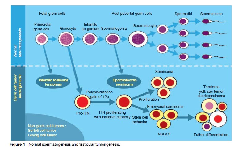
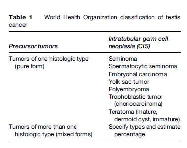
- germ cell tumors (GCT), which account for 94% to 97% of cases, arise from intratubular neoplasia (Figure 1) and are divided into the seminoma and the nonseminomatous germ-cell tumors (NSGCT) (Table 1)
- rare tumors that arise from nongerminal elements (Leydig cells, Sertoli cells, etc.).
In this research paper, I focus on GCT that represent the main public health concern.
Seminoma
Seminoma is the most common GCT type (accounting for 30–50% of GCT). At presentation, the testis is enlarged without loss of normal shape. On macroscopic specimen examination, the tumor is usually solitary with distinct borders. It is pink-white, smooth, and usually homogeneous. Microscopically, seminoma cells are uniform and have clear cytoplasm and well-defined cell borders. Age at presentation is generally several years older than for NSGCT tumors (30–40 vs. 20–35). Three subtypes have been described: typical (85% of the cases), anaplastic (5% to 10%), and spermatocytic (2% to 10%). Initially, the anaplastic variety was thought to carry a worse prognosis than the typical variety; however, currently this difference is debated.
Spermatocytic seminoma usually presents later in the patient’s life. Its prognosis is excellent and cases of metastasis are extremely rare.
Nonseminomatous Germ Cell Tumors
Embryonal carcinoma is an aggressive tumor that tends to metastasize early. Embryonal carcinoma is often associated with other cell types in the metastatic sites. Pure embryonal carcinoma represents 3–6% of GCT.
Choriocarcinoma is another aggressive tumor with a high potential to metastasize (lungs). Even a small intratesticular lesion may present with evidence of advanced distant metastases. Pure choriocarcinoma is extremely rare (1% of GCT), and it is often a component of a mixed tumor.
Teratoma contains more than one germ cell layer in various stages of maturation and differentiation. Mature elements seem like benign structures derived from normal ectoderm, endoderm, and mesoderm; however, in adults, mature teratoma must be considered as malignant because it has the ability to dedifferentiate into more aggressive forms. Immature teratoma consists of undifferentiated primitive tissues from each of the three germ cell layers.
Yolk sac tumor is the most common testis tumor of infants and children. In adults, it occurs almost exclusively in combination with other histological types.
Tumors of more than one histological type are considered a separate entity and are also known as mixed GCT; they make up approximately 60% of all GCTs. Components of embryonal carcinoma and seminoma are very common in mixed GCT.
Incidence
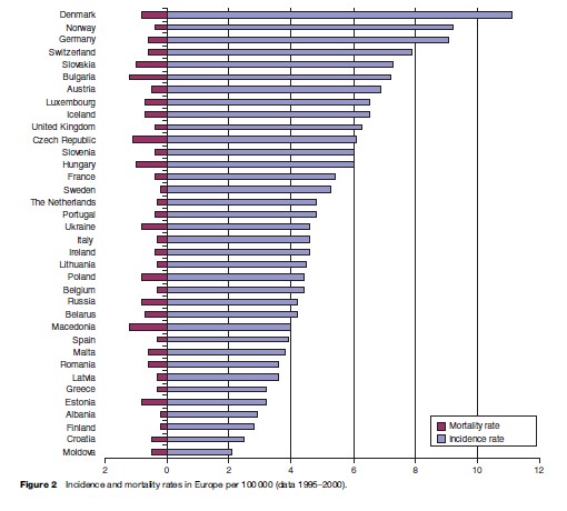
Approximately 500 000 new cases were reported worldwide in 2002. Wide geographical discrepancies exist between the countries: the rates in developed countries are about six times higher than those in developing countries (Huyghe et al., 2003). The present age-standardized incidence rate ranges from less than 1/100 000 in Asia and Africa to up to 10/100 000 in Switzerland and Denmark (Table 1) (Purdue et al., 2005).
Trends In Incidence
Since the Second World War, TC incidence has been increasing in nearly all industrialized countries, especially in Europe, Northern America, and Oceania (Adami et al., 1994; Moller et al., 1995; McGlynn et al., 2003; Richiardi et al., 2004). The worldwide incidence rate of testicular cancer has doubled over the last 50 years. In Europe, strong differences exist between countries (Figure 2) and a gradient in the increase has been observed, the highest rate in Europe is centered in Denmark and Germany, and decreases progressively in a centrifugal way (Huyghe et al., 2006). Recent data may indicate a trend to stabilization in the incidence rate in Denmark, Switzerland, and the United States (Levi et al., 1990; Pharris-Ciurej et al., 1999; Moller, 2001).
Birth Cohort Effect
A clear birth cohort effect (that is, the incidence rate varies with the cohort of birth) has been identified in TC (Bergstrom et al., 1996; Zheng et al., 1996). In Europe, during the last century, the risk of testicular cancer has been increasing regularly with successive birth cohorts; an interruption occurred in this rise in countries such as those in Scandinavia during the Second World War, followed by a new increase after the War (Moller, 1989; Bergstrom et al., 1996). These epidemiological findings have been interpreted to support the hypotheses that TC may be secondary to an early exposure (maybe as early as embryonic or fetal life) to etiologic factors and that these factors may be present in the environment.
Age
Testicular cancer occurs typically in young adults (20 to 35 years), and it is the most frequent malignancy in this age group in several countries. So far the risk of developing TC is predominant in young age groups (15–35 years) (Moller et al., 1995). Overall, the highest incidence is noted in young adult males, making these neoplasms the most common solid tumors of men age 20 to 34 years and the second most common of men age 35 to 40 years. Seminoma is rare before age 10 and after age 60; peak incidence occurs at 35 years. Embryonal carcinoma occurs predominantly between 25 and 35 years, choriocarcinoma more often between 20 and 30 years. Pure yolk sac tumors and teratoma are predominant lesions of childhood but frequently appear in combination with other elements in adulthood.
Racial Factors
The incidence of TC is highest in Europeans and European-derived populations, with age-standardized rates of between 2 to 10 per 100 000 World Standard Population (IARC, 2002; Purdue et al., 2005). In nonEuropean populations the incidence rates are usually below 2 per 100 000. The Maori population of New Zealand is an exception, with an incidence rate of 7 per 100 000 (Wilkinson et al., 1992). Several examples of variation in incidence among ethnic groups come from multiethnic countries: The incidence of TC in American Blacks is approximately one-third that in American Whites (but 10 times that in African Blacks) (McGlynn et al., 2005). In Israel, Jewish people have at least an eight-fold higher incidence than the non-Jewish people (Parkin and Iscovich, 1997). In Hawaii, the incidence in the Filipino and Japanese population is one-tenth that in the white population.
Occupation And Sociodemographic Characteristics
The level of evidence of a link between sociodemographic data and TC incidence rate is low even if higher incidence rates have been noted in the upper and middle socioeconomic classes (Swerdlow et al., 1991).
Significant relationship with the occurrence of TC has been found in men exposed to various environmental or occupational conditions, such as leather tanners, airframe repairers, policemen, gas and petroleum workers, carpet and textile workers, paper, plastic, and metal workers, and pesticide-exposed farmers. However, no definite conclusion can be ascertained, due to the long list of potential causative agents. Regarding the link between occupation of parents and the occurrence of TC in children, TC has been found to be associated with paternal occupations (metalworkers, food and beverage service workers, painter and printing workers, food product workers, wood processors). Interestingly, no relationship was shown with maternal occupations (Pearce et al., 1987; Swerdlow et al., 1991; Van den Eeden et al., 1991; Moller, 1997; Pollan et al., 2001).
Diet
The number of studies examining diet and TC risk is still limited. Intake of milk, dairy products, and cheese has been found to be associated with TC risk by several authors. Cheese and dairy products contain a high level of fat, calcium, and proteins, and they also contain large amounts of the female hormones estrogen and progesterone. The impact of calcium or meat consumption is still debated. Other potential dietary TC risk factors are high saturated, animal, and high total fat intakes and increased intake of baked products (that include milk, sugar, and eggs). The critical periods could be prenatal and perinatal periods, but childhood and adolescence are also pointed out. Relatively late exposures have also been suspected: an association between a high fat consumption 1 year before diagnosis in one study and high dairy product intake two years before diagnosis in another study were associated with an increased risk of TC (Davies et al., 1990, 1996; Bonner et al., 2002).
Laterality
Testicular cancer appears to be slightly more common in the right testis than in the left, similar to the slightly greater incidence of right-sided cryptorchidism. Approximately 2 to 3% of testicular tumors are bilateral, occurring either simultaneously or successively (Sokal et al., 1980). Similar, rather than different, histology in the two testes predominates with bilateral tumors. A history of cryptorchidism (unilateral or bilateral) in nearly half these men is consistent with observations that bilateral dysgenesis occurs frequently in unilateral maldescent. Long-term surveillance of patients with a history of cryptorchidism or previous orchiectomy for GCT is mandatory.
Risk Factors
Cryptorchidism
About 7 to 12% of patients with TC have a prior history of cryptorchidism (the absence of one or both testes from the scrotum) (Schottenfeld et al., 1980; Boyle and Zaridze, 1993). The exact incidence of cryptorchidism is unknown because of difficulties in defining cryptorchidism. Scorer and Farrington estimated that approximately 4.3% of neonates, 0.8% of infants and children, and 0.7% of adults older than 18 years harbor a truly cryptorchid testis (Scorer, 1979). In patients with cryptorchidism, there is an estimated 2 to 32-fold increased risk of TC (Schottenfeld et al., 1980; Herrinton et al., 2003). Between 5% and 10% of patients with a history of cryptorchidism develop malignancy in the controlateral (normally descended) gonad (Berthelsen et al., 1982). The basis for the relationship between cryptorchidism and testicular cancer remains unclear; it is hypothesized that both are different manifestations of a common condition that may result from genetic, lifestyle, and/or environmental factors. Orchiopexy that consists of descending surgically an undescended testis in scrotal position is mandatory in order to preserve fertility (for this purpose, it must be realized early in childhood, preferably before 3 years of age) and to facilitate clinical surveillance of these patients; however, there is no clear evidence that orchiopexy prevents TC (Strader et al., 1988; Jonesl et al., 1991; Cortes et al., 2001; Herrinton et al., 2003; Pettersson et al., 2007).
Hormones
Changes in the testosterone–estrogen balance may contribute to the development of testicular tumor.
Several prenatal risk factors for germ cell cancer may be related with estrogenic disturbances (Moller and Skakkebaek, 1997; Wanderasl et al., 1998; Richiardi et al., 2003):
- Low birth weight due to intrauterine growth retardation
- Low parity of mother
- Children born preterm
- High maternal age of the first-born boy.
Relative risk for testicular tumor in the sons of diethylstilbestrol-treated mothers ranges from 2.8 to 5.3% (Schottenfeld et al., 1980).
Genetic Factors
Although a relatively higher incidence of testicular tumors has been reported in twins, brothers, and family members, genetic causative factors have not been clearly identified. Standardized incidence ratios for testicular cancer are 3.8 and 7.6 when a father and a brother have testicular cancer, respectively (Hemminki and Chen, 2006). However, the high familial risk may be the product of heritable causes or it may also result from shared environmental conditions. Nicholson and Harland (1995) reported that one-third of all testis cancer patients are genetically predisposed to disease, probably a homozygous (recessive) inheritance of a single predisposing gene. Certain genetic conditions are frequently observed in TC (alteration of the sex chromosome isochromosome of 12(i12p), intersex conditions) (Peltomaki, 1991; Peltomaki et al., 1992; Lothe et al., 1995; Bianchi et al., 2002; Smiraglia et al., 2002).
Etiology And Pathogenesis
Experimental and clinical evidence supports the importance of congenital factors in the etiology of GCTs. Existence of a birth cohort effect and data on migrants in Sweden, which show that the relative risk of developing testicular cancer is 0.34 in Finnish migrants to Sweden compared with the Swedish male population, strengthens the hypothesis of a pre and postnatal environmental exposure (Hemminki et al., 2002; Ekbom et al., 2003). The temporal decline of male fertility in humans and wildlife is paralleled by an increase in endocrine-dependent pathologies in the male, such as hypospadias, male infertility, cryptorchidism, and testicular cancer, collectively called Skakkebaek testicular dysgenesis (Skakkebaek et al., 1998, 2001). The ‘estrogen hypothesis’ has been put forward as a possible explanation for these trends. The hypothesis is that in utero, the primordial germ cell may be altered by environmental factors, mainly manufactured substances with estrogen-like action (xenooestrogens) resulting in disturbed differentiation (Skakkebaek, 2002). In vitro studies suggest that some heavy metals also have an estrogen-like action (Chan et al., 1983; Ragan and Mast, 1990). The estrogen-like action seems not to be the only mechanism of action of endocrine disruptors. Some endocrine disruptors are anti-androgenic, such as phthalates (Fisher, 2004). Even if a large body of animal experiments supports the endocrine disruption hypothesis, fewer data are available from human studies (Virtanen et al., 2005; Bay et al., 2006; Frederiksen et al., 2007).
Diagnostic Delay
Patients who present with advanced disease (stage III) generally have a much poorer prognosis than do those with disease confined to the testis or those with regional nodal involvement only. Diagnostic delay, which is defined as the time elapsing from the onset of tumor symptom to the diagnosis of the tumor, has a prognostic value (Huyghe et al., 2007). Diagnostic delay up to six months is not uncommon and seems to be related to survival. It may be correlated to patient factors such as ignorance, denial, and fear, as well as physician factors such as misdiagnosis. As almost half of patients continue to present with metastatic disease, the need clearly exists for population education through campaigns of information on TC symptoms and natural history and programs such as those advocating testicular self-examination (TSE). Only through widespread public health techniques will the knowledge of testicular tumors be promulgated so that diagnosis can occur earlier. Persistent physician-related delay of treatment pleads for continuing education and awareness campaigns. TSE is recommended by the American Medical Association, the American Urological Association, and the American Cancer Society as a regular monthly practice, arguing that TSE is the best way of detecting early TC, especially in men at high risk of developing TC. On the contrary, the United States Preventive Services Task Force and the National Cancer Institute consider that TC screening by TSE would not result in an appreciable decrease in mortality but would lead to numerous unnecessary diagnostic procedures. Although TSE is inexpensive and relatively easy to teach, its acceptance by young males and regular practice remains low in the United States and Europe (Sheley et al., 1991; Singer et al., 1993; Brenner et al., 2003).
Signs And Symptoms
The usual presentation is a lump, painless swelling, or hardness of the testis (60%). Description of an enlargement by patients with a previously small atrophic testis is not uncommon. In approximately 10% of patients, acute pain is the presenting symptom and in 30%, patients may complain of sensation of fullness or heaviness in the inguinal region or scrotum. In a minority of patients (10%), infertility may be the presenting complaint (Raman et al., 2005). On rare occasions (5–10%), patients continue to present with manifestations due to metastases such as a neck mass (supraclavicular lymph node metastasis); respiratory symptoms, such as cough or dyspnea (pulmonary metastasis); gastrointestinal disturbances, such as anorexia, nausea, vomiting, or haemorrhage (retro duodenal metastasis); lumbar back pain (bulky retroperitoneal disease involving the psoas muscle or nerve roots); bone pain (skeletal metastasis); central and peripheral nervous system manifestations (cerebral, spinal cord, or peripheral root involvement); or unilateral or bilateral lower-extremity swelling (iliac or caval venous obstruction or thrombosis). Finally, gynecomastia due to endocrine manifestation of these neoplasms is seen in a majority of Leydig cell tumors and about 5% of patients with testicular GCTs (Gabrilove and Furukawa, 1984; Mellor and McCutchan, 1989).
Physical Examination
Any firm, hard, or fixed area within the substance of the tunica albuginea should be considered suspicious until proved otherwise. In general, seminoma tends to expand within the testis as a painless, rubbery enlargement. Embryonal carcinoma may produce an irregular, rather than discrete, mass, although this distinction is not always easily appreciated.
Testicular tumors tend to remain ovoid (egg-shaped), being limited by the tough investing tunica albuginea. In 10 to 15% of patients, spread to the epididymis or cord may occur. A hydrocele may be present and increase the difficulty of appreciation of a testicular neoplasm. Ultrasonography of the scrotum is a rapid, reliable technique to exclude hydrocele or epididymitis and should be used if there is any suspicion of testicular tumor.
Natural History
All GCT in adults should be regarded as malignant. The only GCT that may be considered as benign is infantile teratoma. Generally, NSGCT are considered as more aggressive than seminoma, and at the time of diagnosis, approximately half of patients with NSGCT present with disseminated disease. Moreover, the growth rate of NSGCT tends to be high. Doubling times range from 10 to 30 days. A total of 85% of patients dying from NSGCT do so within 2 years and the majority of the remainder within 3 years (Aass et al., 1990; Price et al., 1990).
Seminoma has a slower course: approximately 75% of seminomas are confined to the testis at the time of clinical presentation, which represents a smaller percentage than that among patients with NSGCT. Because of this indolent course, seminoma may recur from 2 to 10 years after apparently successful initial management, and a long follow-up is mandatory (Borge et al., 1988; Chung et al., 2002).
Staging
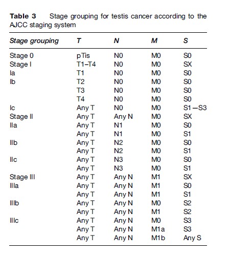
The American Joint Committee on Cancer (AJCC) staging for GCTs conceived a tumor, nodes, metastasis, and serum marker staging (TNMS) system (Tables 2 and 3).
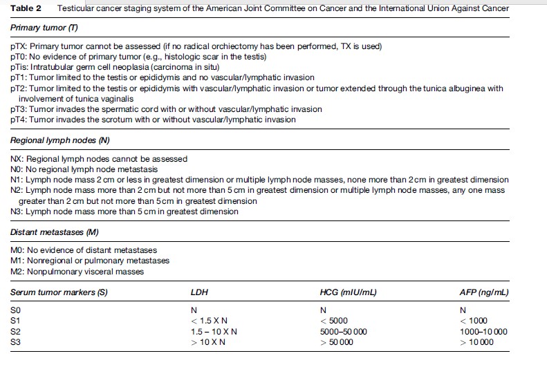
Serum markers used in the AJCC staging are serum human chorionic gonadotropin (HCG), a-fetoprotein (aFP), and lacticodeshydrogenase (LDH).
Treatment
‘Radical’ orchiectomy, consisting of removal of the whole testis by inguinal approach, is the first step of treatment, except in case of bilateral tumor or tumor occurring in a solitary testis that may be treated conservatively. Orchiectomy provides histologic diagnosis local staging (pT) and local control of the neoplasm. Because more than half of patients with testicular tumors present with metastatic disease, further treatment after orchiectomy is usual. Modalities of treatment (surgery, radiotherapy, chemotherapy) depend on histology and staging. Not only multimodal therapy, but also the accuracy of clinical staging and the ability to recognize failure early are of first importance for treatment efficacy.
Prognosis
Seminoma
In stage I seminoma, the 3-year and the 5-year survival rates after treatment are 99% and 95%, respectively. In stages II and III, the 5-year survival rates range from 80 to 90%.
Nonseminomatous Germ Cell Tumor
The cure rate for patients pathologically confirmed stage I NSGCT is roughly 95% with surgery alone. Patients with metastatic NSGCT disease classified as good or intermediate prognosis do well with standard chemotherapy, with response rates of 80–90%. Patients with advanced disease classified as poor prognosis, however, have only a 50–60% therapeutic response. Therefore, more aggressive chemotherapy should be used in this population.
Trends In Mortality
Comparison of specific death rates from testis cancer revealed that in Eastern Europe, mortality decreased slower than in Western Europe, and that decline in mortality occurred later (only since the late 1980s). In the United States, as in Western Europe, mortality has fallen by about 70% since the 1970s as a result of the introduction of modern cisplatin-based chemotherapies.
Bibliography:
- Aareleid T, Sant M, et al. (1998) Improved survival for patients with testicular cancer in Europe since 1978. EUROCARE Working Group. European Journal of Cancer 34(14 Spec. No): 2236–2240.
- Aass N, Fossa SD, et al. (1990) Prognosis in patients with metastatic non-seminomatous testicular cancer. Radiotherapy and Oncology 17(4): 285–292.
- Adami HO, Bergstrom R, et al. (1994) Testicular cancer in nine northern European countries. International Journal of Cancer 59(1): 33–38.
- Bay K, Asklund C, et al. (2006) Testicular dysgenesis syndrome: possible role of endocrine disrupters. Best Practices & Research Clinical Endocrinology & Metabolism 20(1): 77–90.
- Bergstrom R, Adami HO, et al. (1996) Increase in testicular cancer incidence in six European countries: a birth cohort phenomenon. Journal of the National Cancer Institute 88(11): 727–733.
- Berthelsen JG, Skakkebaek NE, et al. (1982) Screening for carcinoma in situ of the contralateral testis in patients with germinal testicular cancer. British Medical Journal (Clinical Research Edition) 285(6356): 1683–1686.
- Bianchi NO, Richard SM, et al. (2002) Mosaic AZF deletions and susceptibility to testicular tumors. Mutatation Research 503(1–2): 51–62.
- Bonner MR, McCann SE, et al. (2002) Dietary factors and the risk of testicular cancer. Nutrition and Cancer 44(1): 35–43.
- Borge N, Fossa SD, et al. (1988) Late recurrence of testicular cancer. Journal of Clinical Oncology 6(8): 1248–1253.
- Boyle P and Zaridze DG (1993) Risk factors for prostate and testicular cancer. European Journal of Cancer 29A(7): 1048–1055.
- Brenner JS, Hergenroeder AC, et al. (2003) Teaching testicular selfexamination: education and practices in pediatric residents. Pediatrics 111(3): e239–e244.
- Chan WY, Chung KW, et al. (1983) Zinc metabolism in testicular feminization and surgical cryptorchid testes in rats. Life Sciences 32(11): 1279–1284.
- Chung P, Parker C, et al. (2002) Surveillance in stage I testicular seminoma – risk of late relapse. Canadian Journal of Urology 9(5): 1637–1640.
- Cortes D, Thorup JM, et al. (2001) Cryptorchidism: aspects of fertility and neoplasms. A study including data of 1,335 consecutive boys who underwent testicular biopsy simultaneously with surgery for cryptorchidism. Hormone Research 55(1): 21–27.
- Davies TW, Prener A, et al. (1990) Body size and cancer of the testis. Acta Oncology 29(3): 287–290.
- Davies TW, Palmer CR, et al. (1996) Adolescent milk, dairy product and fruit consumption and testicular cancer. British Journal of Cancer 74(4): 657–660.
- Ekbom A, Richiardi L, et al. (2003) Age at immigration and duration of stay in relation to risk for testicular cancer among Finnish immigrants in Sweden. Journal of the National Cancer Institute 95(16): 1238–1240.
- Fenner-Crisp PA (2000) Endocrine modulators: risk characterization and assessment. Toxicology and Pathology 28(3): 438–440.
- Fisher JS (2004) Environmental anti-androgens and male reproductive health: focus on phthalates and testicular dysgenesis syndrome. Reproduction 127(3): 305–315.
- Frederiksen H, Skakkebaek NE, et al. (2007) Metabolism of phthalates in humans. Molecular Nutrition and Food Research 51(7): 899–911.
- Gabrilove JL and Furukawa H (1984) Gynecomastia in association with a complex tumor of the testis secreting chorionic gonadotropin: studies on the testicular venous effluent. Journal of Urology 131(2): 348–350.
- Germa-Lluch JR, Garcia del Muro X, et al. (2002) Clinical pattern and therapeutic results achieved in 1490 patients with germ-cell tumors of the testis: the experience of the Spanish Germ-Cell Cancer Group (GG). European Urology 42(6): 553–562; discussion 562–563.
- Hemminki K and Chen B (2006) Familial risks in testicular cancer as aetiological clues. International Journal of Andrology 29(1): 205–210.
- Hemminki K, Li X, et al. (2002) Cancer risks in first-generation immigrants to Sweden. International Journal of Cancer 99(2): 218–228.
- Hendry WF, Norman AR, et al. (2002) Metastatic nonseminomatous germ cell tumors of the testis: results of elective and salvage surgery for patients with residual retroperitoneal masses. Cancer 94(6): 1668–1676.
- Herrinton LJ, Zhao W, et al. (2003) Management of cryptorchism and risk of testicular cancer. American Journal of Epidemiology 157(7): 602–605.
- Huyghe E, Matsuda T, and Thonneau P (2003) Increasing incidence of testicular cancer worldwide: a review. Journal of Urology 170(1): 5–11.
- Huyghe E, Matsuda T, Daudin M, et al. (2004) Fertility after testicular cancer treatments: results of a large multicenter study. Cancer 100(4): 732–737.
- Huyghe E, Plante P, et al. (2006) Testicular cancer variations in time and space in Europe. European Urolology 51(3): 621–628.
- Huyghe E, Muller A, et al. (2007) Impact of diagnostic delay in testis cancer: Results of a large population-based study. European Urolology 52(6): 1710–1716.
- International Agency for Research on Cancer (IARC) (2002) Cancer Incidence in Five Continents vol. 8. Lyon, France: IARC Scientific Publication.
- Joly F, Heron JF, et al. (2002) Quality of life in long-term survivors of testicular cancer: a population-based case-control study. Journal of Clinical Oncology 20(1): 73–80.
- Jones BJ, Thornhill JA, et al. (1991) Influence of prior orchiopexy on stage and prognosis of testicular cancer. European Urology 19(3): 201–203.
- Levi F, Te VC, et al. (1990) Changes in cancer incidence in the Swiss Canton of Vaud, 1978–87. Annals of Oncology 1(4): 293–297.
- Lothe RA, Peltomaki P, et al. (1995) Molecular genetic changes in human male germ cell tumors. Laboratory Investigations 73(5): 606–614.
- McGlynn KA, Devesa SS, et al. (2003) Trends in the incidence of testicular germ cell tumors in the United States. Cancer 97(1): 63–70.
- McGlynn KA, Devesa SS, et al. (2005) Increasing incidence of testicular germ cell tumors among black men in the United States. Journal of Clinical Oncology 23(24): 5757–5761.
- Mellor SG and McCutchan JD (1989) Gynaecomastia and occult Leydig cell tumor of the testis. British Journal of Urology 63(4): 420–422.
- Moller H (1989) Decreased testicular cancer risk in men born in wartime. Journal of National Cancer Institute 81(21): 1668–1669.
- Moller H (1997) Work in agriculture, childhood residence, nitrate exposure, and testicular cancer risk: a case-control study in Denmark. Cancer Epidemiology Biomarkers & Prevention 6(2): 141–144.
- Moller H (2001) Trends in incidence of testicular cancer and prostate cancer in Denmark. Human Reproduction 16(5): 1007–1011.
- Moller H, Jorgensen N, et al. (1995) Trends in incidence of testicular cancer in boys and adolescent men. International Journal of Cancer 61(6): 761–764.
- Moller H and Skakkebaek NE (1997) Testicular cancer and cryptorchidism in relation to prenatal factors: Case-control studies in Denmark. Cancer Causes Control 8(6): 904–912.
- Mostofi FK, Davis J Jr., et al. (1994) Tumours of the Testis. Lyon, France: IARC Scientific Publication (111): 407–429
- Mostofi FK and Sesterhenn IA (1985) Pathology of germ cell tumors of testes. Progress in Clinical and Biological Research 203: 1–34.
- Moul JW, Paulson DF, et al. (1990) Delay in diagnosis and survival in testicular cancer: impact of effective therapy and changes during 18 years. Journal of Urology 143(3): 520–523.
- Nicholson PW and Harland SJ (1995) Inheritance and testicular cancer. British Journal of Cancer 71(2): 421–426.
- Parkin DM and Iscovich J (1997) Risk of cancer in migrants and their descendants in Israel: II. Carcinomas and germ-cell tumours. International Journal of Cancer 70(6): 654–660.
- Pearce N, Sheppard RA, et al. (1987) Time trends and occupational differences in cancer of the testis in New Zealand. Cancer 59(9): 1677–1682.
- Peltomaki P (1991) DNA methylation changes in human testicular cancer. Biochimica et Biophysica ACTA 1096(3): 187–196.
- Peltomaki P, Lothe RA, et al. (1992) Chromosome 12 in human testicular cancer: dosage changes and their parental origin. Cancer Genetics and Cytogenetics 64(1): 21–26.
- Pettersson A, Richiardi L, et al. (2007) Age at surgery for undescended testis and risk of testicular cancer. New England Journal of Medicine 356(18): 1835–1841.
- Pharris-Ciurej ND, Cook LS, et al. (1999) Incidence of testicular cancer in the United States: has the epidemic begun to abate? American Journal of Epidemiology 150(1): 45–46.
- Pollan M, Gustavsson P, et al. (2001) Incidence of testicular cancer and occupation among Swedish men gainfully employed in 1970. Annals of Epidemiology 11(8): 554–562.
- Post GJ and Belis JA (1980) Delayed presentation of testicular tumors. Southern Medical Journal 73(1): 33–35.
- Price P, Hogan SJ, et al. (1990) The growth rate of metastatic nonseminomatous germ cell testicular tumours measured by marker production doubling time–II. Prognostic significance in patients treated by chemotherapy. European Journal of Cancer 26(4): 453–457.
- Purdue MP, Devesa SS, et al. (2005) International patterns and trends in testis cancer incidence. International Journal of Cancer 115(5): 822–827.
- Ragan HA and Mast TJ (1990) Cadmium inhalation and male reproductive toxicity. Review of Environmental Contamination and Toxicology 114: 1–22.
- Raman JD, Nobert CF, et al. (2005) Increased incidence of testicular cancer in men presenting with infertility and abnormal semen analysis. Journal of Urology 174(5): 1819–1822; discussion 1822.
- Richiardi L, Askling J, et al. (2003) Body size at birth and adulthood and the risk for germ-cell testicular cancer. Cancer Epidemiology Biomarkers & Prevention 12(7): 669–673.
- Richiardi L, Bellocco R, et al. (2004) Testicular cancer incidence in eight northern European countries: secular and recent trends. Cancer Epidemiology Biomarkers & Prevention 13(12): 2157–2166.
- Schottenfeld D, Warshauer ME, et al. (1980) The epidemiology of testicular cancer in young adults. American Journal of Epidemiology 112(2): 232–246.
- Scorer CG (1979) Cryptorchidism: a renewed plea. British Medical Journal 1(6163): 616.
- Sheley JP, Kinchen EW, et al. (1991) Limited impact of testicular selfexamination promotion. Journal of Community Health 16(2): 117–124.
- Singer AJ, Tichler T, et al. (1993) Testicular carcinoma: A study of knowledge, awareness, and practice of testicular self-examination in male soldiers and military physicians. Military Medicine 158(10): 640–643.
- Skakkebaek NE (2002) Endocrine disrupters and testicular dysgenesis syndrome. Hormone Research 57(Suppl 2): 43.
- Skakkebaek NE, Rajpert-De Meyts E, et al. (1998) Germ cell cancer and disorders of spermatogenesis: an environmental connection? Acta Pathologica Microbiologica et Immunologica Scandanavica 106(1): 3–11; discussion 12.
- Skakkebaek NE, Rajpert-De Meyts E, et al. (2001) Testicular dysgenesis syndrome: an increasingly common developmental disorder with environmental aspects. Human Reproduction 16(5): 972–978.
- Smiraglia DJ, Szymanska J, et al. (2002) Distinct epigenetic phenotypes in seminomatous and nonseminomatous testicular germ cell tumors. Oncogene 21(24): 3909–3916.
- Sokal M, Peckham MJ, et al. (1980) Bilateral germ cell tumours of the testis. British Journal of Urology 52(2): 158–162.
- Strader CH, Weiss NS, et al. (1988) Cryptorchism, orchiopexy, and the risk of testicular cancer. American Journal of Epidemiology 127(5): 1013–1018.
- Swerdlow AJ, Douglas AJ, et al. (1991) Cancer of the testis, socioeconomic status, and occupation. British Journal of Industrial Medicine 48(10): 670–674.
- Van den Eeden SK, Weiss NS, et al. (1991) Occupation and the occurrence of testicular cancer. American Journal of Industrial Medicine 19(3): 327–337.
- Virtanen HE, Rajpert-De Meyts E, et al. (2005) Testicular dysgenesis syndrome and the development and occurrence of male reproductive disorders. Toxicology and Applied Pharmacology 207(2 Suppl): 501–505.
- Wanderas EH, Grotmol T, et al. (1998) Maternal health and pre and perinatal characteristics in the etiology of testicular cancer: a prospective population and register-based study on Norwegian males born between 1967 and 1995. Cancer Causes & Control 9(5): 475–486.
- Wilkinson TJ, Colls BM, et al. (1992) Increased incidence of germ cell testicular cancer in New Zealand Maoris. British Journal of Cancer 65 (5): 769–771.
- Zheng T, Holford TR, et al. (1996) Continuing increase in incidence of germ-cell testis cancer in young adults: experience from Connecticut, USA, 1935–1992. International Journal of Cancer 65(6): 723–729.
- Boisen KA, Kaleva M, Main KM, et al. (2004) Difference in prevalence of congenital cryptorchidism in infants between two Nordic countries. Lancet 363(9417): 1264–1269.
- Bosl GJ and Motzer RJ (1997) Testicular germ-cell cancer. New England Journal of Medicine 337(4): 242–253.
- Bray F, Richiardi L, Ekbom A, Pukkala E, Cuninkova M, and Moller H (2006) Trends in testicular cancer incidence and mortality in 22 European countries: Continuing increases in incidence and declines in mortality. International Journal of Cancer 118(12): 3099–3111.
- Brenner H (2002) Long-term survival rates of cancer patients achieved by the end of the 20th century: A period analysis. Lancet 360(9340): 1131–1135.
- Herrinton LJ, Zhao W, and Husson G (2003) Management of cryptorchism, risk of testicular cancer. American Journal of Epidemiolology 157(7): 602–605.
- Horwich A, Shipley J, and Huddart R (2006) Testicular germ-cell cancer. Lancet 367(9512): 754–765.
- Levi F, LaVecchia C, Boyle P, Lucchini F, and Negri E (2001) Western, eastern European trends in testicular cancer mortality. Lancet 357 (9271): 1853–1854.
- Logothetis CJ (1988) Adjuvant chemotherapy for testicular cancer. New England Journal of Medicine 319(1): 53.
- Oliver RT (2001) Trends in testicular cancer. Lancet 358(9284): 841.
- Oliver RT, Mason MD, Mead GM, et al. (2005) Radiotherapy versus single-dose carboplatin in adjuvant treatment of stage I seminoma: a randomised trial. Lancet 366(9482): 293–300.
- Pharris-Ciurej ND, Cook LS, and Weiss NS (1999) Incidence of testicular cancer in the United States: has the epidemic begun to abate? American Journal of Epidemiology 150(1): 45–46.
- Wein AJ, Kavoussi LR, Novick AC, Partin AW, and Peters CA (2007) Campbell – Walsh Urology, 9th edn. Philadelphia, PA: Saunders, Elsevier.
See also:
Free research papers are not written to satisfy your specific instructions. You can use our professional writing services to buy a custom research paper on any topic and get your high quality paper at affordable price.


